Fetal heart rate (FHR) testing is a primary method used to determine the health of the fetus before birth and to help identify potential risks such as hypoxia or stress in the fetus. The purpose of early detection is to reduce fetal morbidity and mortality.
Currently, the most common method of fetal heart rate detection is Doppler ultrasound, and the standard prenatal fetal health test is the fetal no-load test (NST). These tests are usually performed in a hospital with continuous wave instruments.
Although the current ultrasonic fetal heart rate tester has been greatly improved, the price is decreasing, and the volume is smaller, we still need accurate sensor calibration and certain expertise to operate the detector correctly. In addition, such instruments are quite sensitive to movement, and the safety issues that may result from long-term exposure to ultrasound in the fetus are not yet conclusive. Therefore, the use of the detector is now limited to short-term testing.
Another method of measuring fetal heart rate is fetal electrocardiogram (FECG), but the steps are more complicated and less practical. Moreover, commercial non-invasive FECG devices have not yet appeared on the market.
Recently, an optical method which is still in the research stage has been proposed, which uses a halogen lamp or a tungsten lamp as a light source to realize detection by photomultiplier. However, these techniques are costly, require high light intensity, and are difficult to implement due to instrument size and power consumption limitations.
Optical fetal heart rate detection systemOur research team proposed a low-power optical technique based on photoplethysmogram (PPG) signals to non-invasively detect fetal heart rate. The PPG signal is generated by the pulsation of light through the blood. The doctor or technician uses an LED light (less than 68 mW) to illuminate the abdomen of the pregnant woman, and the beam is modulated by the blood circulation of the mother and the fetus. The maximum wavelength of light that can be penetrated is 890 nm. The mixed signal can be analyzed by adaptive filtering using digital signal processing, and the pregnant woman's index finger PPG is used as a reference input.
Develop an optical fetal heart rate (OFHR) inspection system using LabVIEW graphical system design software and NI hardware. In the OFHR system, the SNR decreases as the incident power decreases; the excitation signal is the modulated beam. The system can implement synchronous detection. The software subroutine in LabVIEW uses the NI 9474 digital output module to generate the modulation frequency on the counter.
At the receiver, low noise amplification and synchronous detection ensure that the useful information is saved with minimal noise power. The 24-bit NI USB-9239 analog-to-digital converter (ADC) reduces the effects of quantization noise. Once digitization is complete, the signal is processed by an adaptive noise canceller (ANC) technique to extract the fetal PPG from the mixed signal.
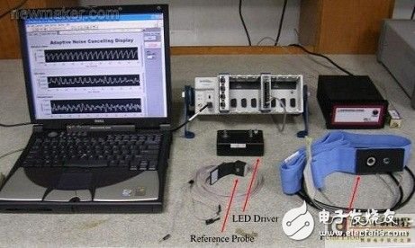
Use a belt to connect the fetal probe (primary signal) to the abdomen of the pregnant woman, keeping the IR-LED at a distance of 4 cm from the photodetector. Connect the reference probe to the mother's index finger. Since the selected IR-LED can only emit a maximum power of 68 mW, the operating optical power of the OFHR system is set to be less than the 87 mW specified by the International Commission on Non-Ionizing Radiation Protection (ICNIRP). To modulate the IR-LED, a software subroutine is used to generate a 725 Hz modulated signal that is connected to the LED driver via the NI 9474 counter terminal (Figure 1). In Figure 1, the diffuse reflected light from the abdomen of the pregnant woman is measured by a low noise photodetector and is expressed in the form of I (M1, F), where M1 and F represent the effects of the mother's abdomen and the fetus on the signal, respectively.

Figure 1: The hardware block in the OFHR system block diagram is implemented by the LabVIEW program. A low noise (6 nV/Hz1/2) transimpedance amplifier converts the current into a voltage. The reference probe (connected to the mother's index finger) consists of an IR-LED and a solid-state photodiode with a built-in preamplifier. The signal from the probe is denoted as I (M2); M2 represents the effect of the mother on the signal. This channel does not require simultaneous detection because the photoplethysmogram of the index finger has a high signal-to-noise ratio (SNR).
The NI USB-9239 24-bit resolution data acquisition module simultaneously acquires signals from both probes at a rate of 5.5 kHz. Demodulation, signal filtering, and signal estimation are performed in the digital domain. The software implements modulation signal generation, synchronization detection algorithms, downsampling, high-pass filtering, and adaptive noise cancellation (ANC) algorithms.
The design team used LabVIEW to implement the entire algorithm and some of the instruments. After completing the pre-processing and application of the ANC algorithm, LabVIEW will display the fetal signal and fetal heart rate results.
Figure 2a shows the laboratory prototype and graphical user interface of the OFHR system, and gives the pregnant index finger PPG (top), abdominal PPG (middle), and the estimated PPG of the fetus (bottom).

Figure 2a: OFHR prototype
Figure 2b shows three optional displays, including digital sync or lock-in amplifier (LIA), adaptive noise canceller (ANC), and heart rhythm trajectory. The first two displays are available for assisted development and the third is used to indicate values ​​for fetal heart rate versus time. Users can view the data online or save it for further analysis.
Figure 2b: Graphical user interface of the OFHR system
After the completion of the development, we tested the functionality of the system based on a total of 24 sets of data from 6 clinical subjects ranging from 35 to 39 weeks of pregnancy, data provided by the National University of Malaysia Medical Center. All fetuses participating in the study were examined by an obstetrician and were healthy without complications.
In the study, we obtained a correlation coefficient of 0.97 between optical and ultrasonic fetal heart rate (p value less than 0.001) with a maximum error of 4%. Clinical results show that the closer the probe is to the fetal tissue (not limited to the brain or buttocks), the better the signal quality and detection accuracy.
in conclusionThe research team developed a new OFHR detection system using low-cost, low-power IR lamps and commercially available silicon detectors. By using LabVIEW, we can quickly and easily implement digital sync detection and adaptive filtering techniques. The accuracy of the measured fetal heart rate results is higher compared to the standard measurement method (Doppler ultrasound). Based on the novelty of the program, we are currently applying for patents in its commercial field.
Stylus Pen For Microsoft Surface
Product catagories of Stylus Pen For Microsoft Surface, which just can be worked on below Surface model, Please confirm your surface model before buying.
Microsoft Surface 3; Microsoft Surface Pro 3; Microsoft Surface Pro 4; Microsoft Surface Pro 5; Microsoft Surface Pro 6; Microsoft Surface Book; Microsoft Surface Laptop; Microsoft Surface Studio.
Stylus Pen For Microsoft Surface,Tablet Touch Pen,Touch Screen Stylus Pen,Universal Stylus Pen
Shenzhen Ruidian Technology CO., Ltd , https://www.wisonens.com