At the time of infection, the initial CD8+ T cells were activated, proliferated and differentiated into effector CD8+ T cells, with an increase in body temperature. To know whether elevated body temperature will further regulate the activation and differentiation of CD8+ T cells, we must first solve the quantitative analysis of morphological changes during T cell activation and differentiation.
Amnis' ImageStream high-content microscopic imaging flow cytometer combines the advantages of fluorescence microscopy and flow cytometry to obtain images of each cell while rapidly analyzing large numbers of cells for objective quantitative analysis. We know that when the fever is happening, the body is resisting foreign pathogens. In the past, we only knew that high temperature can inhibit the replication ability of pathogens. But recent studies by the Immunization and Flow Cytometry team at the Rozwell Park Cancer Institute (RPCI) in New York, USA have shown that fever can also improve the function of the immune system, specifically CD8+ T cells. Function [Thomas A. Mace, JLB, 2011].
In this study, Amnis's ImageStream high-content microscopic imaging flow cytometer played an invaluable role, not only perfectly solving the technical bottleneck of traditional flow analysis and traditional image observation, but also its powerful image. Quantitative analysis capabilities also make it a tool for immunological research. Inflammation or infection by pathogens, the initial CD8+ T cells are activated by interaction with APC cells, which in turn increase and differentiate into effector CD8+ T cells, which may be accompanied by an increase in body temperature. However, people still do not understand whether the increase in body temperature will further regulate the activation and differentiation of CD8+ T cells.
To solve this problem, the first thing to solve is how to quantitatively analyze the morphological changes during the activation and differentiation of T cells. The ImageStream high-content microscopic imaging flow cytometer from Amnis in the United States solves this problem: the system combines the advantages of fluorescence microscopy and flow cytometry to obtain a large number of cells while simultaneously obtaining each cell. Images and objective, quantitative analysis of these images.
Recently, the results of their study were published in the Journal of White Blood Biology of the Rozwell Park Cancer Institute in New York, USA: The researchers used ImageStream high-content microscopic imaging flow cytometry for lower body temperature (33 ° C and The binding of mouse CD8+ T cells to APC cells at 37 ° C) and higher body temperature (39.5 ° C) was quantitatively analyzed. The results showed that the increase in body temperature increased the binding rate of the two.
More importantly, the researchers also found that the number of CD8+ T cells in the experimental group increased significantly, and the GM1 and CD8 co-receptors on the membrane surface aggregated into clusters, which affected the fluidity of the membrane. These results reveal that an increase in body temperature caused by fever can affect the stress effects regulated by antigen-specific CD8+ T cells by increasing the number of effector cell populations. In this study, ImageStream showed its powerful image quantitative analysis capabilities.
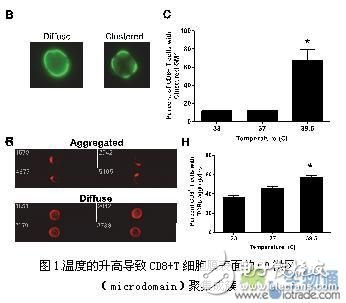
The researchers first isolated the original CD8+ T cells from the experimental mice and then analyzed the fluidity of the cell membranes by ImageStream after incubation at 33 ° C, 37 ° C, and 39.5 ° C for 6 hours. Since ImageStream can image the detected cells, it can be visualized to show that the FITC-labeled GM1 protein is evenly distributed or clustered (Fig. 1B).

Quantitative image analysis of whole population cells by software, the researchers used the BrightDetail Intensity parameter in ImageStream software to analyze the cells, and found that the GM1 clustering on the cell membrane surface increased significantly after the temperature increased to 39.5 °C (Fig. 1C). In addition, it has been reported that the TCR signal complex also clusters on the surface of the cell membrane when T cells are activated. Therefore, the researchers also analyzed the complex and similar results were obtained (Fig. 1G and Fig. H).
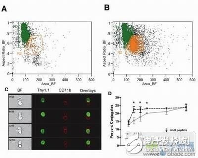
To determine whether temperature would affect the formation of CD8+ T cells and APC cells, the researchers incubated the primary CD8+ T cells at 37° or 39.5°C for 6 hours and then co-cultured with C57BL/6 spleen cells. The group was treated with gp10025-33 polypeptide. Finally, ImageStream was used to analyze the number of secondary syncytial cells that developed immune synapses.
First, distinguish between single-cell and two-connected cells by Mingfield and Aspect RaTIo (short axis/long axis of cells) (Fig. 2A and B, R1 is a single cell, R2 is a binary cell), and then pass CD8+Thy1. The expression of .1+ (ie CD8+ T cells) and CD11b+ (ie APC) was gated to select for T cell and APC duplex cells (Fig. 2C). The experimental results show that the increase in temperature causes a significant increase in the ratio of CD8+ T cells to APC spliced ​​cells (Fig. 2D).

Figure 2. Increase in temperature leads to an increase in the proportion of dimers in CD8+ T cells and APC cells.
Next, the researchers hope to explore whether the co-receptors of CD8+ T cells will also aggregate into clusters as the temperature increases. This co-receptor molecule, like TCRβ, is a very important molecule for CD8+ T cell activation. The author's guess was confirmed by research (Figures 3A and 3B).

Figure 3. Elevation of mice leads to an increase in the proportion of co-receptor clusters on the surface of CD8+ T cell membranes.
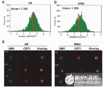
At the same time, the researchers also used the Similarity parameter in ImageStream software to analyze the co-localization of CD8 co-receptor and GM1 in the cell membrane, but the results showed that there was no difference in the similarity value before and after the increase in body temperature of the mice (data not shown). To further investigate the clustering of GM1 in T cells, the investigators analyzed the colocalization of GM1 and the negative control CD71, and the results showed a decrease in the value of similarity before and after the increase in body temperature in mice (right panels C and D). The images show that CD71 molecules are not co-clustered with GM1 after elevated body temperature in mice, as shown in Figure E on the right.

Finally, the researchers believe that CD8+ T cells and their differentiation into effector cells are temperature sensitive. If CD8+ T cells are at a higher temperature first, then they will respond more quickly and efficiently when they receive antigenic stimulation. In this type of immunology research, the biggest technical bottleneck encountered by researchers is how to quantify a large number of cell images and obtain statistically significant data—because traditional flow cytometry cannot obtain morphological information of cells. The use of fluorescence microscopy requires human judgment and analysis, and the number of cells counted is very limited. The ImageStream high-content microscopic imaging flow cytometer launched by Amnis in the United States perfectly solves this problem, enabling rapid and objective protein translocation, molecular colocalization, morphological changes, molecular endocytosis, immune synapse formation, etc. Morphology was quantitatively analyzed.
Samsung Screen Protector,Tempered Glass Samsung,Mobile Tempered Glass,Tempered Glass Screen Protector Iphone Samsung
Shenzhen TUOLI Electronic Technology Co., Ltd. , https://www.hydrogelprotector.com